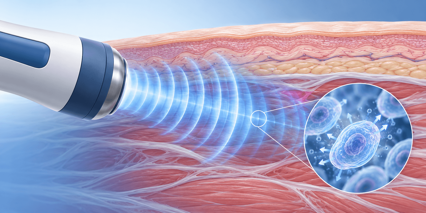
From Acoustic Energy to Tissue Interaction
A shockwave carries energy as a pressure transient. When it enters tissue, it encounters interfaces between materials of different acoustic impedance—tendon, bone, fascia, muscle, fluid—and the wave is partly reflected, partly transmitted, and partly converted into mechanical stress. That stress appears as tension, compression, and shear at the cellular and extracellular matrix level.
This is a physical description, not a biological one. The mechanical disturbance is the input; the biological response is the output. The gap between them is where mechanotransduction begins.
What Is Mechanotransduction?
Mechanotransduction is the process by which cells convert mechanical forces into intracellular biochemical signals. Cells possess mechanosensors—integrins at focal adhesions, stretch-activated ion channels, purinergic receptors, and cytoskeletal components—that detect deformation and initiate signaling.
For shockwave therapy specifically, distinct experimental lines have identified purinergic signaling as one candidate mechanism. However, these studies use different cell types and different receptor subsets, and should not be merged into a single confirmed universal pathway.
Cellular and Molecular Responses Under Investigation
Cell Signaling and Tissue Remodeling
In a 2025 in vitro study, Szwarc-Hofbauer and colleagues used C3H/10T1/2 mouse mesenchymal stromal cells to examine purinergic signaling after shockwave exposure. They reported that P2Y2 and P2X4 receptors are primarily expressed in these cells, and that shockwave treatment induced c-Jun and c-Fos expression within 30 minutes. Erk1/2 activation and immediate early gene expression were decreased in P2Y2-, P2X4-, and double-deficient cells. Shockwave treatment did not promote proliferation in double-knockout cells, while loss of either single receptor significantly reduced the proliferative effect, indicating a cumulative contribution. This is a mouse cell line studied in vitro; it establishes a receptor-specific mechanism in that system, not a human tendon or bone response.
A separate study used human bone marrow–derived mesenchymal stem cells. Sun et al. (2013) reported that shockwave treatment released approximately 7 μM ATP, activated P2X7 receptors and p38 MAPK, and induced osteogenic differentiation; blocking any of these steps prevented differentiation. This is a different cell type (primary human MSCs), a different receptor (P2X7), a different kinase (p38), and a different outcome (osteogenic differentiation, not proliferation).
These two studies are parallel, cell-type-specific findings. They do not establish a unified ATP → purinergic receptor → MAPK → proliferation/differentiation cascade across tissues.
Other laboratory work has implicated integrin-mediated MAPK/ERK and Wnt/β-catenin signaling in osteogenic responses, and has described matrix remodeling through collagen synthesis and matrix metalloproteinase regulation. These findings remain largely in vitro and animal-based.
Angiogenesis and Vascular Responses
Neovascularization is among the more consistently reported biologic effects. In a dog model, low-energy shockwave application (1,000 impulses at 14 kV, approximately 0.18 mJ/mm²) to the Achilles tendon–bone junction increased new capillaries and muscularized vessels at 4 and 8 weeks, with none in control specimens. In vitro and animal studies also report upregulation of vascular endothelial growth factor (VEGF) and endothelial nitric oxide synthase (eNOS) after shockwave exposure.
Angiogenesis is a plausible contributor to repair, but it does not by itself establish clinical improvement for a given indication.
Focused Shockwaves and Radial Pressure Waves: Mechanistic Differences
Both focused shockwaves (FSW) and radial pressure waves (RPW) are applied transcutaneously and are described as producing mechanotransduction. The mechanistic difference lies primarily in energy delivery geometry.
A focused shockwave converges toward a defined depth, with peak pressure at a target determined by device design, applicator, and tissue density. A radial pressure wave is generated at the applicator surface and propagates divergently, with maximal energy near the tip and dissipation as it travels outward. Cells in a deeper focal volume therefore experience different pressure transients than cells in more superficial tissue exposed to a decaying wave.
What this means for downstream biology is less settled. Some evidence suggests FSW can promote osteoprogenitor differentiation and address bone-related conditions, while RPW is used primarily for tendinopathies and fasciopathies. Comparative mechanistic literature is not robust enough to claim that one modality produces a categorically different set of cellular responses across all tissues. Delivery geometry differs; the biological consequences of that difference are indication- and parameter-dependent.
What Laboratory Studies Can—and Cannot—Tell Us
Laboratory studies answer a narrow question: under specified conditions, does shockwave exposure produce a measurable cellular or molecular change? They identify candidate mechanisms and generate hypotheses.
They cannot answer whether a patient will experience less pain or recover function. Cell culture systems lack the architecture, vascular supply, immune context, and mechanical loading of living tissue. Animal models are closer, but species differences, injury models, and protocol variations limit direct translation. The ISMST/DIGEST Conjoint Physics Working Group has noted that currently established shockwave parameters used to describe in vitro or clinical conditions are insufficient, and that the key effect of shockwaves on tissue is not yet fully known.
Human clinical evidence is indication-specific. A mechanism demonstrated in a mouse cell line or a canine tendon does not establish a human clinical effect.
How Treatment Parameters Influence Biological Responses
Energy flux density, pulse count, frequency, and coupling technique all affect the mechanical dose delivered to tissue. Different parameter sets can produce different cellular responses in laboratory models—and in some cases, opposing responses depending on energy level and cell type.
Biological response is dose- and context-dependent. A protocol validated for one device, energy level, and indication should not be assumed to produce the same mechanobiological effect when any of those variables change.
Conclusion
Shockwave therapy works, at the most basic level, by delivering mechanical stress that cells convert into biochemical signals. Mechanotransduction is the entry point; downstream effects under investigation include changes in cell proliferation and differentiation, angiogenesis, matrix remodeling, inflammatory signaling, and pain pathways. The strength of evidence differs at each layer: physical principles are established; receptor-level mechanisms are cell-type- and model-specific; human clinical efficacy is indication-specific.
For how these mechanisms fit into clinical decision-making and U.S. For the evidence behind benefits and side effects, see Shockwave Therapy Benefits / Side Effects.
Frequently Asked Questions
Does mechanotransduction fully explain how shockwave therapy works?
No. Mechanotransduction describes the initial conversion of mechanical force into cellular signaling. It does not by itself explain why some patients respond and others do not, or why clinical results vary across indications. Multiple downstream pathways—inflammatory, vascular, neural, and matrix-related—are under investigation, and their relative contribution in human tissue is not fully mapped.
If a laboratory study shows cell proliferation or angiogenesis, does that mean shockwave therapy will heal the tissue?
Not necessarily. Laboratory findings establish that a biological response is possible under controlled conditions. They do not establish clinical efficacy in a specific patient population. Angiogenesis and cell proliferation are plausible contributors to repair, but they are not substitutes for clinical outcome data.
Do focused and radial devices work through the same biological mechanism?
Both are described as producing mechanotransduction, but they deliver different mechanical environments. Focused waves converge at depth; radial waves diverge from the applicator surface. The downstream biology may overlap, but the comparative evidence is not strong enough to claim identical mechanisms or interchangeable effects across all tissues.
Why do different studies report different mechanisms?
Because shockwave therapy is not one intervention. Energy level, pulse count, frequency, device type, target tissue, and measurement timing vary. A pathway activated in one cell line at one energy level may not be activated in another model at a different energy level.
References
- 1. Szwarc-Hofbauer D, Simböck E, Hromada C, Stoiber M, Tomasch J, Weitzer G, Teuschl-Woller AH. Purinergic receptors play a key role in shock wave-induced proliferation. Scientific Reports. 2025;15:19138. doi:10.1038/s41598-025-02955-3. Source View source
- 2. Sun D, Junger WG, Yuan C, et al. Shockwaves induce osteogenic differentiation of human mesenchymal stem cells through ATP release and activation of P2X7 receptors. Stem Cells. 2013;31(6):1170–1180. doi:10.1002/stem.1356. Source View source
- 3. Wang CJ, Huang HY, Pai CH. Shock wave-enhanced neovascularization at the tendon-bone junction: an experiment in dogs. Journal of Foot and Ankle Surgery. 2002;41(1):16–22. doi:10.1016/S1067-2516(02)80005-9. Source View source
- 4. International Society for Medical Shockwave Treatment (ISMST). ESWT Guidelines, English version, 3rd edition. Daegu, 2023. Mechanism of action chapter, pp. 17–19. Source
- 5. Guo Z, Yuan Y, Su H, Dong J. Extracorporeal shock wave therapy in knee osteoarthritis: a mechanistic narrative review of neurogenic inflammation and the MMP/ADAMTS axis. International Journal of General Medicine. 2026;19:629190. doi:10.2147/IJGM.S629190. Source View source
This article is educational and does not replace individualized clinical assessment, current device instructions, or applicable regulatory labeling.